


Ongoing Event in United States of America from Jul 26 to Jul 30, 2026
Innovative Solutions for Automated Imaging
MetaSystems has been developing and producing system software for automated microscopy since 1986. Today, users in more than 103 countries rely on the laboratory solutions by MetaSystems.
In addition to generating the highest quality images, our modern approaches include an advanced workflow management that grows with your requirements.
You would like to discover what others are accomplishing with their MetaSystems products? Read through the list of peer-reviewed publications from our customers!
If you do not find what you are looking for, please do not hesitate to contact the MetaSystems partner in your region.
RENEB Intercomparison Exercises Analyzing Micronuclei
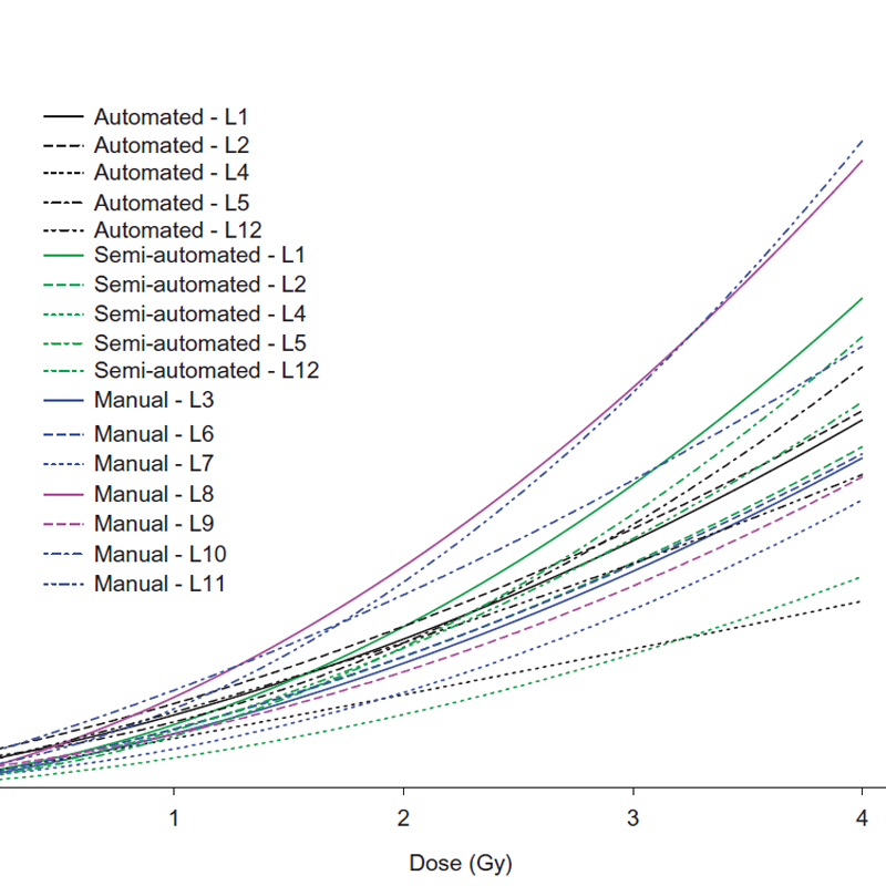
Radiological emergencies in which a large number of victims are involved can occur as the result of different events such as radiation accidents, terrorist attacks with dirty bombs or other kinds of exposure to radioactive sources. In the event of large radiological accidents, an initial triage aimed at classifying victims according to the degree of exposure is indicated to guide appropriate clinical responses. [...] The cytokinesis-block (CB) micronucleus (MN) assay, originally developed by Fenech and Morley in 1985, is a valuable biodosimetric tool for quantifying radiation-induced chromosomal damage for population triage thanks to the simplicity of MN scoring and the availability of automated MN analysis using microscopy-based [...] methods. [...] Five laboratories performed both automated and semi-automated MN scoring using the Metafer platform (Metasystems, Altlussheim, Germany). [...] The lowest level of inhomogeneity was obtained for the automated scoring group where a more standardized approach was applied. [...] The results of the two micronucleus intercomparison exercises performed by the RENEB network, demonstrate that the cytokinesis-block MN assay is a useful triage tool for large-scale
radiation emergencies.
Cited From: International journal of radiation biology (2017)
Read more...
All Publications
Related Products/Solutions
Are You Looking for Probes?
![]() MetaSystems Probes now has its own website. If you are looking for MetaSystems XCyting DNA Probes, please visit the MetaSystems Probes website.
MetaSystems Probes now has its own website. If you are looking for MetaSystems XCyting DNA Probes, please visit the MetaSystems Probes website.
Next Event
Latest News
Advancing Laboratory Automation in Argentina

A New Chapter for MetaSystems
The MetaSystems Foundation (MetaSystems-Stiftung), registered as a non-profit organization, aims to preserve the founders’ life’s work while providing employees with stable jobs and long-term development opportunities in the region.
