


Ongoing Event in United States of America from Jun 28 to Jul 2, 2026
Innovative Solutions for Automated Imaging
MetaSystems has been developing and producing system software for automated microscopy since 1986. Today, users in more than 103 countries rely on the laboratory solutions by MetaSystems.
In addition to generating the highest quality images, our modern approaches include an advanced workflow management that grows with your requirements.
You would like to discover what others are accomplishing with their MetaSystems products? Read through the list of peer-reviewed publications from our customers!
If you do not find what you are looking for, please do not hesitate to contact the MetaSystems partner in your region.
Slide Imaging and Image Analysis for Diatom Morphometrics
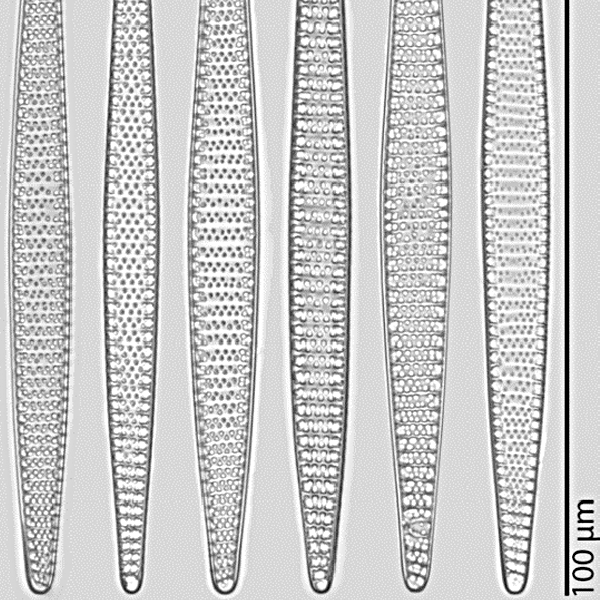
Light microscopy analysis of diatom frustules is widely used in basic and applied research [...]. For image acquisition, we use a Metafer slide scanning system [...]. In position list
mode, a list of user-defined positions, spread arbitrarily over the slide, can be imaged. [...] The vertical position of objects can be determined by automatic creation of a focus map, i.e., based on interpolation between reference points, or by autofocusing at each position captured, or using a combination of both. [...] Our two-cycle microscopy and image analysis procedure starts with a low-resolution area scan [...]. The overlapping low-resolution images are stitched together to a virtual slide using the VSlide software [...]. For locating and selecting valves of interest, the low-resolution images [...] are processed by SHERPA. [...] We use the VSViewer software to import the position list provided by SHERPA [...] into the virtual slide [...]. The annotation function of the viewer enables to manually add positions of interest if some objects deemed relevant were missed by SHERPA [...]. This allows for combining (semi-)automated and manual selection of valves of interest. [...] The annotated virtual slide from step 5 contains the (X, Y) position data required for selectively imaging the valves of interest. [...]. Extending the size range reported for F. kerguelensis (quite substantially, by nearly 45% in the case of apical length) nicely illustrates the advantages of a (semi-)automated approach formicroscopic morphometry of diatom valves, primarily that it makes documenting and measuring large numbers of specimens feasible with moderate effort.
Cited From: Appl. Sci. (2017)
Read more...
All Publications
Related Products/Solutions
Are You Looking for Probes?
![]() MetaSystems Probes now has its own website. If you are looking for MetaSystems XCyting DNA Probes, please visit the MetaSystems Probes website.
MetaSystems Probes now has its own website. If you are looking for MetaSystems XCyting DNA Probes, please visit the MetaSystems Probes website.
Next Event
Latest News
MetaSystems Installation for Sperm Detection

Successful completion of the TEAM research project: New approaches to personalized therapy for severe infections
