


Blood Cell Detection
AI-enhanced Cell Classification for Hematological Testing
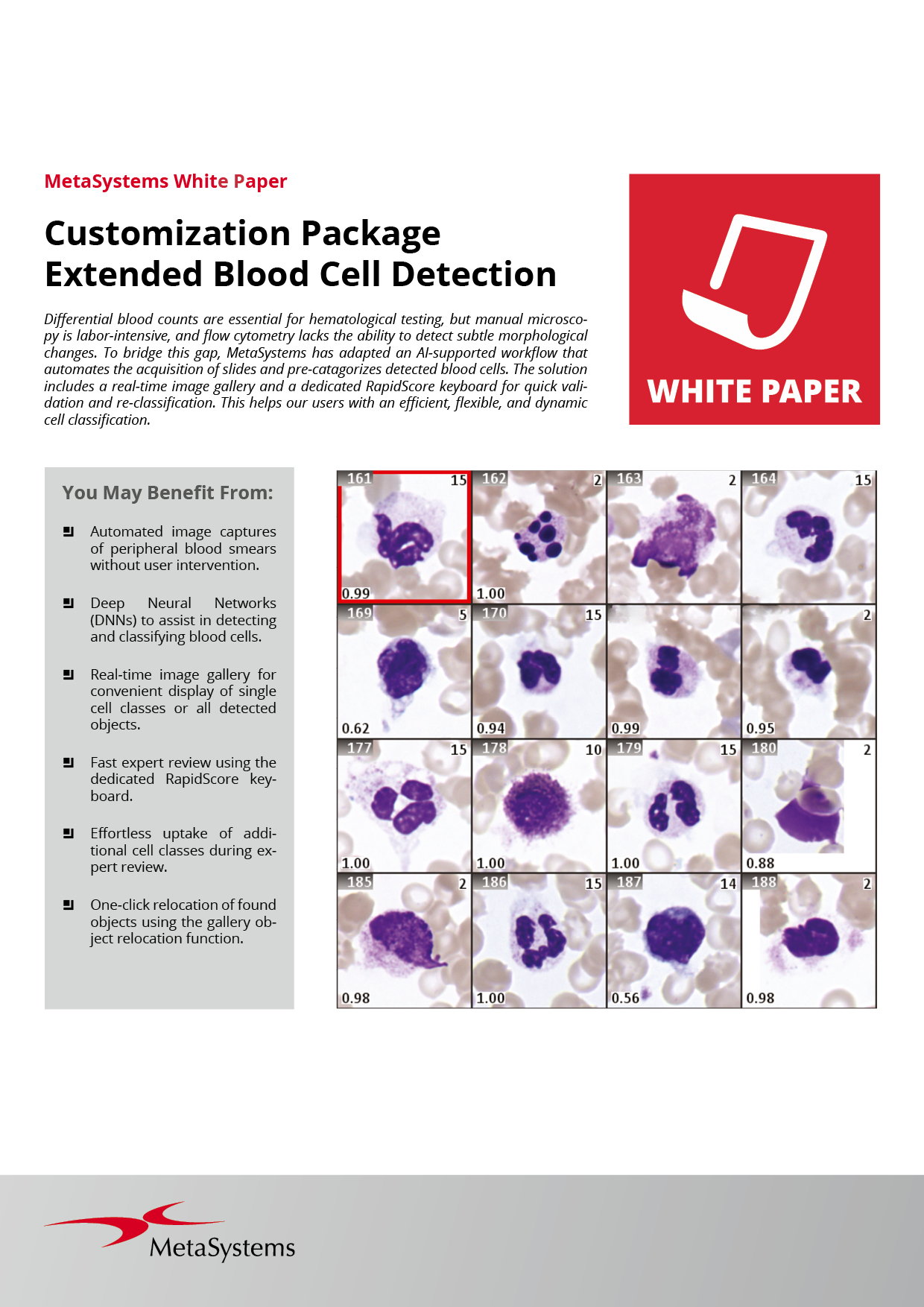
You may benefit most from the following customizations:
- Automated image captures of peripheral blood smears without user intervention.
- Deep Neural Networks (DNNs) to assist in detecting and classifying blood cells.
- Real-time image gallery for convenient display of single cell classes or all detected objects.
- Fast expert review using the dedicated RapidScore keyboard.
- Effortlessly uptake of additional cell classes during review.
- One-click relocation of found objects.
- Scalable to process up to 800 slides in a single run.
- Efficient management of cases and images with Neon.
Differential blood counts are a cornerstone in hematological diagnostics, used to identify and monitor leukemia, inflammatory responses, and infections. While automated systems based on flow cytometry are limited in their ability to detect subtle morphological abnormalities, manual microscopy is time-consuming and labor-intensive.
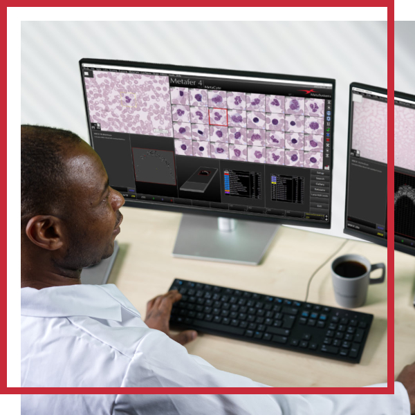
To address this gap, we configured a powerful AI-powered workflow in close collaboration with our users. This workflow enables automatic acquisition of Romanowsky-stained slides and preclassification of blood cells.
The software offers a real-time image gallery that conveniently displays either selected cell classes or all detected objects, supporting an efficient and structured workflow. For fast expert review, the dedicated RapidScore keyboard enables quick validation and reclassification of pre-identified cells. During the review process, additional cell classes can be effortlessly included, allowing users to adapt the classification dynamically and with maximum flexibility.
Looking for Collaboration Partners
We are committed to working at the forefront of novel technologies. This enables us to provide our users with cutting-edge tools for their work. For many of our customers, Deep Neural Networks (DNN) are a powerful tool for further customizing their workflows to their needs. We are currently looking for collaboration partners to help us classify images of bone marrow smears that serve as training data for the networks. Would you like to learn more? Contact us!
The implemented AI can identify single-cell layer areas and supports various slide preparation methods and staining techniques.
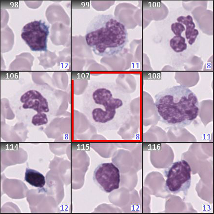
A Deep Neural Network (DNN) trained on expert-annotated images pre-classifies the detected blood cells into 16 categories (state as of May 2025): Artifacts, Nucleated RBCs (incl. Normoblasts), Myeloblasts, Promyelocytes, Myelocytes, Meta-Myelocytes, Band Granulocytes, Segmented Granulocytes, Basophil Granulocytes, Eosinophil Granulocytes, Monocytes, Lymphocytes, Atypical lymphocytes (incl. Reactive Lymphocytes, Neoplastic Lymphocytes), Plasma cells, Smudge cells, and Platelets. These classifications are suggestions provided by the AI and require confirmation or correction by a trained operator.
While RBCs are not further pre-characterized, stored FOV images assist users in evaluating RBC morphology. Customized calculations and queries can be incorporated to further tailor the workflow to user needs.
We understand that relocating individual cells using the eyepiece is crucial in critical cases. That is why the software allows you to view a previously detected object on the slide with a single click. Metafer supports reliable cell relocation even after de-staining the slide for subsequent FISH analysis, enabling you to view the same cell side by side.
RapidScore is a crucial step in the workflow that allows experts to efficiently evaluate pre-classified objects. This process combines automation and human insight to draw well-informed conclusions from the images. The RapidScore keyboard displays the different categories on its keys, allowing experts to swiftly confirm or modify the software-generated proposals with a single keystroke.
Every lab operates differently, and the time needed to process a slide depends on the sample area on the slide and the customized scanning process. This means that we cannot make general statements in this regard. With the support of our application specialists, you can optimize the settings for image acquisition and modify them to suit your individual needs.
- Metafer (6.3MB)
Metafer combines automation, precision, and scalability for modern labs. It controls microscope features like contrasting modes, objectives, and filters, while intelligent auto-focusing ensures sharp images without manual adjustments. Scalable and flexible, Metafer supports the SlideFeeder x80 for up to 800 slides, an automatic oil dispenser for smooth magnification changes, and a barcode reader for efficient high-throughput scanning.
Also available in: - CP Extended Blood Cell Detection (403.8kB)
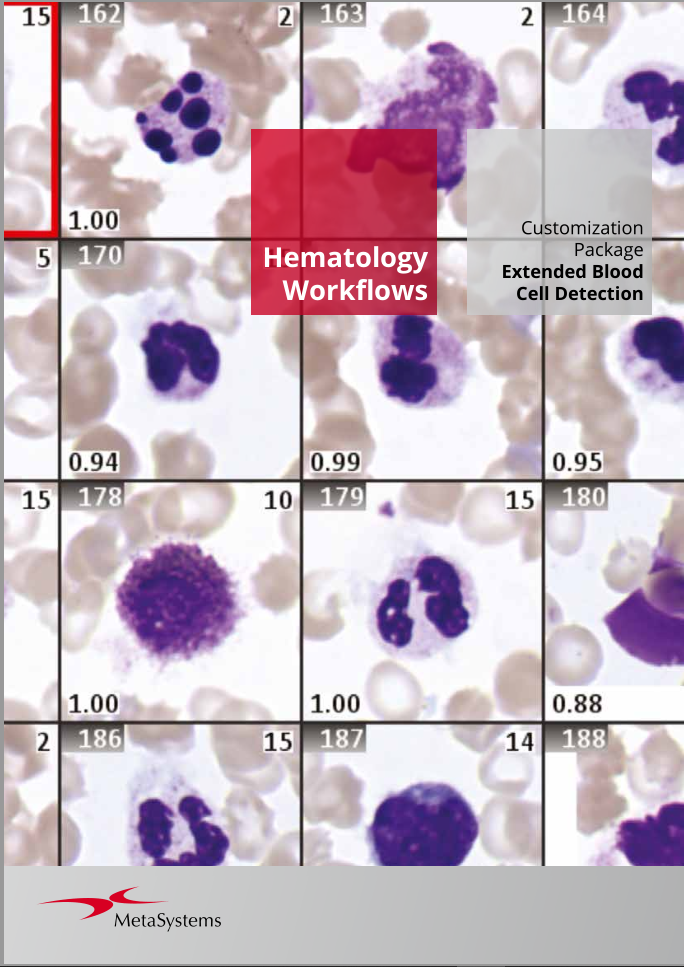
Whole Blood Cell (WBC) counting determines the relative distribution of leukocyte subtypes and is crucial for diagnosing infections, immune disorders, and hematological diseases. The Customization Package for Extended Blood Cell Detection enables you to use DNN-powered workflows for automated whole blood cell counting with your Metafer installation. The workflows integrate DNNs trained on expert-annotated data and are ready for implementation and validation in your lab.
Also available in: - White Paper: Customization Package Extended Blood Cell Detection (6.4MB)
MetaSystems has adapted an AI-supported workflow that automates the acquisition of slides and pre-catagorizes detected blood cells. The solution includes a real-time image gallery and a dedicated RapidScore keyboard for quick validation and re-classification.
Also available in:
MetaSystems software provides, among other functions, features to assist users with image processing. These include, but are not limited to, the use of machine and deep learning algorithms for pattern recognition. The output generated in this process should be regarded as preliminary suggestions and, in any case, mandatorily requires review and assessment by trained experts.
MetaSystems offers Customization Packages for application workflows that have been successfully implemented for customer labs using standard Metafer platform functionality. It is expected that they can be implemented for other customer labs using similar workflows and slide preparation procedures. If a Customization Package is purchased, MetaSystems product specialists will – based on their experience from other similar application cases - support the customer lab in adapting the Metafer software configuration to their needs. The performance of the solution will depend on the quality of the customer slides and the expertise of the users, MetaSystems cannot specify or guarantee any performance parameters. The validation of the solution for clinical use is the sole responsibility of the customer lab.