


Successful completion of the TEAM research project: New approaches to personalized therapy for severe infections
Infective endocarditis is a life-threatening biofilm infection of the inner linings of the heart, including the heart valves. Often, the heart valves are damaged so severely that they must be replaced. Patients receive antibiotic therapy before and after surgery, and timely and precise pathogen diagnosis is essential for the success of this treatment.
However, antibiotic therapy has so far been tailored only with regard to pathogen species and resistance. This approach is particularly inadequate for biofilm-associated infections, as antibiotics often fail in these cases. This leads to “empirical” therapy, the success of which is frequently limited.
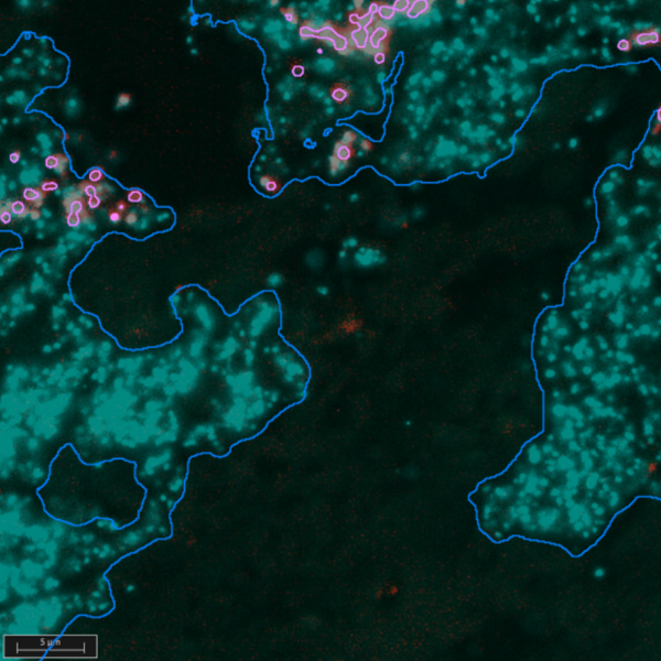
To improve diagnosis and treatment, the TEAM research project—[1]—was launched with funding from the Federal Ministry of Research, Technology, and Space (BMFTR). Using the photonic method of “fluorescence in situ hybridization” (FISH), the foundations for tailored therapy were laid by developing diagnostic and digital procedures capable of analyzing severe bacterial infections of the heart more precisely and, based on these results, guiding antibiotic therapies in a more targeted manner. With the help of FISH, bacteria in biofilms within tissue sections of removed heart valves can be visualized, identified, and their number and activity determined.
Project partners included MoKi Analytics GmbH, Leipzig (project coordination, Dr. Judith Kikhney), the Institute of Medical Microbiology and Virology at Leipzig University Hospital (Prof. Dr. Annette Moter), Intavis Peptide Services GmbH, Tübingen (Dr. Steffen Hüttner), MetaSystems Hard & Software GmbH, Altlussheim (Dr. Andreas Plesch), and NEXUS / CHILI GmbH, Dossenheim (Dr. Uwe Engelmann).
After a four-year duration, the project has now been successfully completed.
“This severe, often bacterial inflammation of the heart’s inner lining or valves remains a disease with a high mortality rate despite all advances. Our goal was to visualize the underlying microbiological processes directly on clinical material and thus better individualize treatment decisions,” explains Prof. Dr. Annette Moter.
At the core of the development is a so-called biofilm classifier, which combines modern molecular biological methods with automated image analysis and artificial intelligence. Innovative PNA-FISH probes were developed for this purpose.
“With our specific PNA-FISH probes, we can visualize microbial activity with significantly greater sensitivity than before. This is an important prerequisite for better understanding and specifically treating infections,” says Dr. Steffen Hüttner of Intavis Peptide Services.
The FISH signals (or markers) are captured and evaluated using automated microscopy. “The combination of automated image capture and AI analysis enables, for the first time, an objective and reproducible assessment of the infection status and the success of preoperative antibiotic therapy as a basis for postoperative infection treatment,” explains Dr. Andreas Plesch of MetaSystems.
“We were able to demonstrate that new approaches for postoperative risk assessment and treatment decisions can be derived from the data. In the future, this will allow us to support physicians in a much more targeted manner,” adds Prof. Dr. Annette Moter.
A central component of the project was the development of a multimodal FISH archive (or PACS: Picture Archive and Communication System) by NEXUS / CHILI GmbH for the structured management and analysis of the resulting image data. This integrates microscopic images with macroscopic images of the collected samples and makes them available within a shared context.
“We consolidate all imaging information related to a sample into a single system and make its interconnections visible. This creates, for the first time, a seamless digital process chain from the sample to the treatment decision,” says Dr. Uwe Engelmann.
“The concepts developed are not limited to a single specialty. We also see great potential in fields such as virology or pathology,” emphasizes Dr. Judith Kikhney, project coordinator at MoKi Analytics.
After the project’s completion, the plan is to further develop the demonstrators into a market-ready medical device that opens up new possibilities for digital analysis and documentation, particularly for diagnostic laboratories using FISH technology.
With the successful completion of TEAM, an important step has been taken toward data-driven, personalized infectious disease medicine—with the potential to sustainably improve diagnostics, treatment planning, and clinical decision-making processes.
The TEAM collaborative project was funded by the Federal Ministry of Research, Technology, and Space (BMFTR) as part of the Photonics Research Germany funding program under grant numbers 13N15815–13N15820.
[1] https://www.quantensysteme.info/projektatlas/projekte/q/team